Instruments
For paraffin embedding:
- Please label cassettes in pencil
- Place tissue into cassettes and close cassettes securely. Please make sure the tissue is small enough to move within the cassette (no thicker than a nickel). If it is too large you might need to dissect it into smaller pieces for it to fix properly. Hard objects such as sutures, hair, or lens will affect sectioning and if not the object of the study, need to be removed prior to fixation/ tissue submission to the core. If tissue is very small, please use biopsy cassettes with denser mesh and add sponges to hold tissue in place.
- Tissue needs to be fixed immediately. Place cassettes into a container large enough to allow the cassettes to move around and fill it with 10% neutral buffered formalin (10%NBF) or 4%PFA. Please use at least 15-20 volumes of formalin for every volume of tissue.
- Fix tissues at room temperature for 24-48hr.
- After fixation, remove formalin and replace it with 70% ethanol.
- Bring your samples to the Histology core in 70% ethanol in a SEALED container (The core no longer accepts samples in inappropriate containers). If you don’t have the right container, please ask us, and we will try to help you find one.
For Preparing Frozen Samples:
- Select a proper size cryomold and label it with permanent marker. Choose a mold larger than your samples. Having enough OCT around the tissue allows for better sample attachment to the chuck and makes sectioning easier. If your sample is sticking out of the mold, the Core staff won’t be able to section it.
- If your sample is in PBS or some other liquid, try to blot away as much of the liquid as possible before placing tissue in OCT. Equilibrate your sample to OCT at room temperature for 30 seconds in a small container (i.e. 50ml tube cap) before moving it to the molds (lungs need to be perfused with a 1:1 mixture of OCT and PBS to remove air before freezing to ensure good section quality and correct tissue morphology).
- Add a few drops of OCT to the mold, transfer your sample and orient it correctly in the mold. The side of the sample facing the bottom of the cryomold is the side that will get sectioned first. Tissue should be placed in the center of the mold. If embedding several pieces of tissue in the same block, place them as close to the center as possible.
- If needed, carefully add more OCT to completely cover the specimen. Avoid bubbles.
- Using long forceps, place the mold in vapors of liquid nitrogen/dry ice-isopentane slurry and allow OCT to freeze completely. It should take 30 sec-1 min for OCT to harden and become white. It is preferred that fresh samples are frozen in liquid nitrogen vapors.
- Some samples might require fixation before freezing in OCT. Samples that are pre-fixed should be incubated in 15% sucrose in PBS, and then in 30% sucrose in PBS until they sink in 30% sucrose. Sucrose helps to displace water and to prevent ice crystals formation inside the sample, which results in a better tissue morphology. Pre-fixed samples can be frozen on dry ice.
- Wrap each mold in aluminum foil to prevent samples from drying and write the sample ID on the foil using permanent marker. Place the block in a labeled zip lock bag. Store samples at -80C and transport them on dry ice.
For Slide Scanning:
- All samples must be clearly labeled, have a coverslip that completely covers the sample, and must be completely dry before submitting to the facility. If using an aqueous-based mounting media, seal the slides with nail polish.
- Protected Health Information (clinical resection number, patient’s name, ect) is NOT allowed on slide labels and must be covered with a new flat and thin label with a coded sample ID.
- All slides must be clean. Depending on the type of the mounting media used, use xylene, 70% ethanol, or commercial slides/objectives cleaning solution to remove mounting media residues, fingerprints and dust from the top and the bottom surfaces of the slides.
- Try your best to avoid bubbles on the sample. Regions adjacent to bubbles are likely to be out of focus.
- If preparing fluorescently labeled samples, avoid using mounting media with DAPI. Staining for DAPI separately before putting a coverslip on always produces better results. We recommend ProLong Diamond Antifade Mountant from Molecular Probes.
- Broken slides can sometimes be manually scanned on the Aperio in brightfield but cannot be scanned on the Vectra.
Please use the following link to place an order with RHTIC in iLab:
- Bring samples to RHTIC together with the printed iLabs-generated order form (please bring histology samples to MSB E311 and samples for scanning and analysis to COMRB 6094).
- Please allow at least 10 business days (although it can take longer depending on the queue) for your order to be processed. If you need it sooner, please let us know – there will be an additional RUSH charge applied for the expedited service
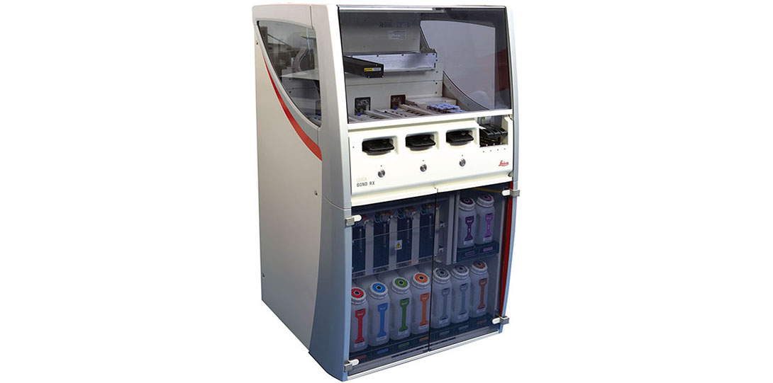
BOND RX Immunostainer Heading link
The Bond RX is a fully automated IHC and ISH staining platform for biotechnology and pharmaceutical research. The Bond RX can be used for standard and dual IHC, multi target IF, ACDBio RNAScope and PerkinElmer Opal Multiplex IHC. It has a 30 slide capacity in three trays.
Microm International GmbH HM340 E Microtom Heading link
The Microm HM340 E is a multi-purpose microtome for use with paraffin, hard- and semithin-sectioning techniques. It will cut sections from .5 µm to 100 µm thick.
Leica LMD7000 Laser Capture Microdissection Heading link
Laser Microdissection (LCM) is a contact- and contamination-free method for isolating specific single cells or entire areas of tissue from a wide variety of tissue samples. The LCM is located in the College of Dentistry.
Leica RM2155 Microtom Heading link
The Leica RM 2155 microtome is fully motorized and can cut sections from 0.5 to 60 microns with an automatic coarse feed. It features an adjustable hand wheel for speed control and selectable cutting modes. The knife holder comes with new clamping plates, ensuring the user accurate sections.
Leica RM2255 Microtom Heading link
The Leica RM2125 is a rotary microtome for paraffin sectioning. Its specimen orientation system has an anti-tilt function that ensures accurate orientation of the specimen surface relative to the knife. The RM2125 universal knife holder base operates with a lateral displacement feature so that the full length of the knife edge can be used without having to change the tension setting on the previously clamped knife or blade.
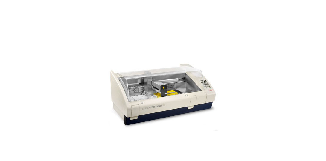
Leica Biosystems ST5010 Autostainer XL Heading link
The Leica Autostainer XL is an efficient H&E and special stain autostainer which can stain up to 200 slides per hour. There are 18 reagent stations 5 wash stations and an integrated forced hot air oven that significantly reduces slide drying time. It allows for simultaneous staining of up to 11 racks (30 slides/rack) using several different protocols. It has continuous load/unload drawer access and robot arm system which allows users to continue slide processing without interrupting the instrument operation. The programming is flexible such that it allows a stain protocol to begin or end in any station and move in any station sequence that maximizes utilization of the instrument while also not allowing a stain protocol to begin that is incompatible with a protocol already in progress.
Leica CV5030 Glass Coverslipper Heading link
The Leica CV5030 is a fully automated glass coverslipper which produces slides with superior optical quality for reliable long-term storage.
Thermofisher Microm HM525 Cryo Heading link
The Microm HM 525 cryostat is used for sectioning of frozen specimens. The chamber will go from 5°C down to –35°C. It will create sections from 1 to 10 microns in 1 micron steps and 11-20 microns in 2 microns steps. Trimming thickness can be from 10-500 microns.
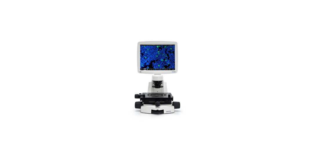
ThermoFisher Evos FL Heading link
The ThermoFisher Evos FL is an inverted digital microscope for fluorescent and transmitted light applications. It has an LED light source and a phase contrast 4x objective and non-phase 10x, 20x, 40x and an oil 100x objective. It can accept most types of plates and slides for imaging. It has Dapi (Ex 357/ Em 477), GFP (Ex 470/ Em 525) and RFP (Ex 531/ Em 593) filters for fluorescence.
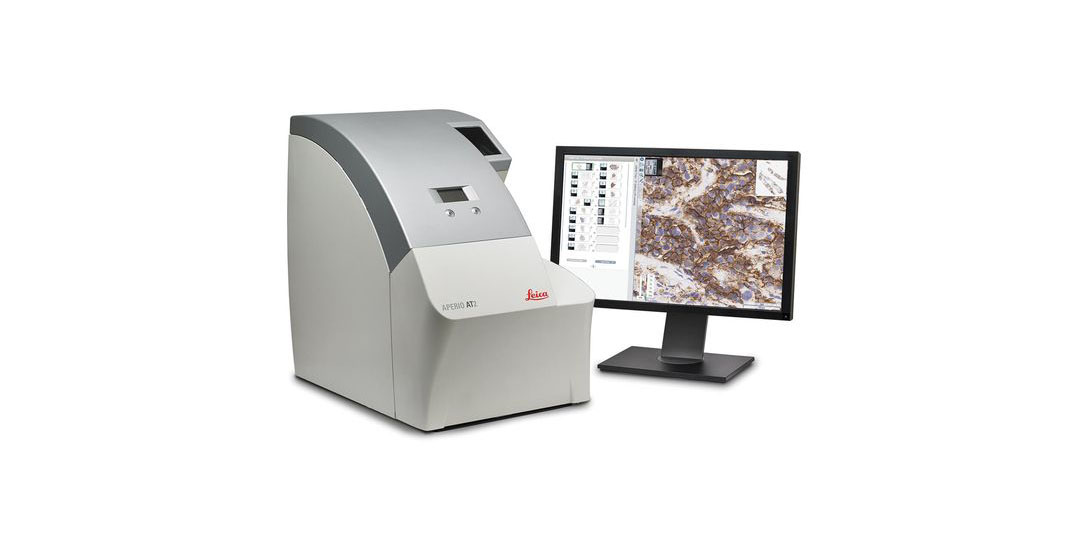
Leica Aperio AT2 Heading link
The Leica Aperio AT2 is a whole slide brightfield scanner for 1″ x 3″ standard microscope slides including whole sections, biopsies and tissue microarrays. The Aperio can scan slides at either 5x, 20x or 40x magnification.
PerkinElmer Vectra 3 Heading link
The PerkinElmer Vectra 3 is a multi-spectral imaging system for use in both brightfield and fluorescence applications. The Vectra requires that samples be prepared on standard 1″ x 3″ microscope slides. Samples can be cultured cells, cell spreads, tissue sections or tissue microarrays. The Vectra has 4x, 10x, 20x and 40x objectives. Whole slide scanning using the Vectra is possible however the instrument is designed to randomly sample tissue of interest at high power for quantification.
Leica MZ16 Dissecting Microscope Heading link
The Leica MZ16 fluorescence stereomicroscope can be used to view and image three-dimensional objects. The instrument has magnifications from 7.1x to 230x and has Dapi and FITC filters for fluorescence viewing. The camera is a SPOT RT KE attached to a computer with SPOT and SPOT advanced software for image acquisition.
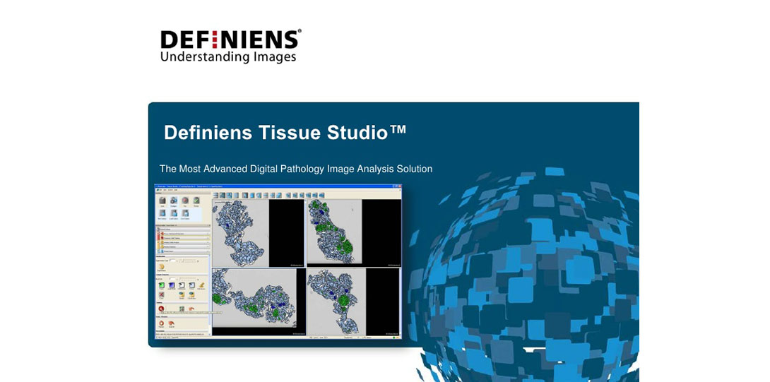
Definiens Tissue Studio Heading link
Tissue Studio is a flexible image analysis platform that allows for quantification of a wide variety of image types. Some examples include single and dual chromegen IHC, up to 3 channel fluorescence quantification, CISH, FISH and tissue microarrays. Definiens can score images acquired from a variety of sources including Leica Aperio, Hamamatsu Nanozoomer and the Zeiss LSM Confocal microscopes.
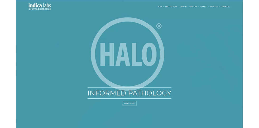
Indica Labs Halo Heading link
Halo is a modular suite of software tools for annotating and analyzing digital images that are either brightfield or fluorescent in origin. The annotation tools are advanced and provide for concentric and tiled ROI partitioning, line segment division, margin creation, automatic layer thickness calculations and whole slide image registration. It can analyze fields of view, whole slides or tissue microarrays and provide subcellular data per individual cell. Halo can process images from either the Leica Aperio AT2 or PerkinElmer Vectra3 as well as a wide variety of whole slide scanners and standard image formats.