Instruments
Instrument time can be scheduled on iLabs. Please check the Getting Started page to register on iLabs.
ZEISS LASER SCANNING MICROSCOPES: LSM 710 META and LSM 710 BIG
Members of the LSM 710 confocal microscope family are popular, full-featured instruments capable of optically sectioning living or fixed specimens of varying thickness. The specialized META detector can be used to capture detailed spectral information and customize emission filters for the elimination of signal overlap. An intuitive time-lapse imaging function is useful for kinetic studies and FRAP experiments, and the laser configuration settings are suitable for FRET measurements. Though fundamentally similar to the LSM 510 META capabilities, the more advanced LSM 710 META confocal microscope provides greater sensitivity and reduced background noise during acquisition. The upgraded ZEN software and “Smart Setup” feature allows for easy importing of fluorophore configurations, while the Definite Focus component can help to correct axial drift during long-term capture. The LSM 710 BIG is equipped with environmental controls (for temperature and CO2) and with two GaAsP detectors (BiG) allowing measurements of very low laser levels, therefore enabling the confocal to be used for long term live cell experiments.
ZEISS TOTAL INTERNAL REFLECTION FLUORESCENCE (TIRF) MICROSCOPE
The versatile TIRF microscope is configured for several types of imaging, most notably for the precise visualization of structures or processes near cell membranes (< 200 nm). The system is also uniquely suited for live cell studies by way of a fully controllable (temperature, CO2, humidity) environmental chamber, which can supplement any of the transmitted light, fluorescence, or laser TIRF imaging modes.
OLYMPUS VIVAVIEW INCUBATOR/FLUORESCENCE MICROSCOPE
The VivaView Fluorescence Microscope system consists of a motorized inverted microscope that is fully contained within a cell culture incubator. This integrated approach to long-term time-lapse imaging can thus extend the duration of acquisition, reduce thermal drift, and allow for precise chemical loading while providing high quality fluorescent and/or transmitted light capture. The system also features a motorized stage for monitoring multiple positions in a dish, as well as an automated tray with space for up to 8 dishes.
OLYMPUS BX51 FLUORESCENCE MICROSCOPE (Upright)
The BX51 fluorescence microscope is a simple fluorescence and bright field instrument with an assortment of objectives ranging from 4X to 100X magnification, as well as a set of 4 manual sliding fluorescence filters (DAPI, FITC, Rhodamine, Cy5). The easy setup and highly-responsive CCD camera make the system an excellent choice when observations and analyses require a certain efficiency.
Prairie Technologies Ultima In Vivo Microscopy System
The Prairie Technologies Ultima In Vivo Microscopy System, 2-Photon imaging is designed specifically for intravital preparations, with fully motorized control of the objective X-Y-Z position. The system is configurable to wide range of live samples from invertebrates to primates. A second scan path enables simultaneous photoactivation or photobleaching. A high-speed resonant scanner brings video-rate imaging to the platform.
slideshow Heading link
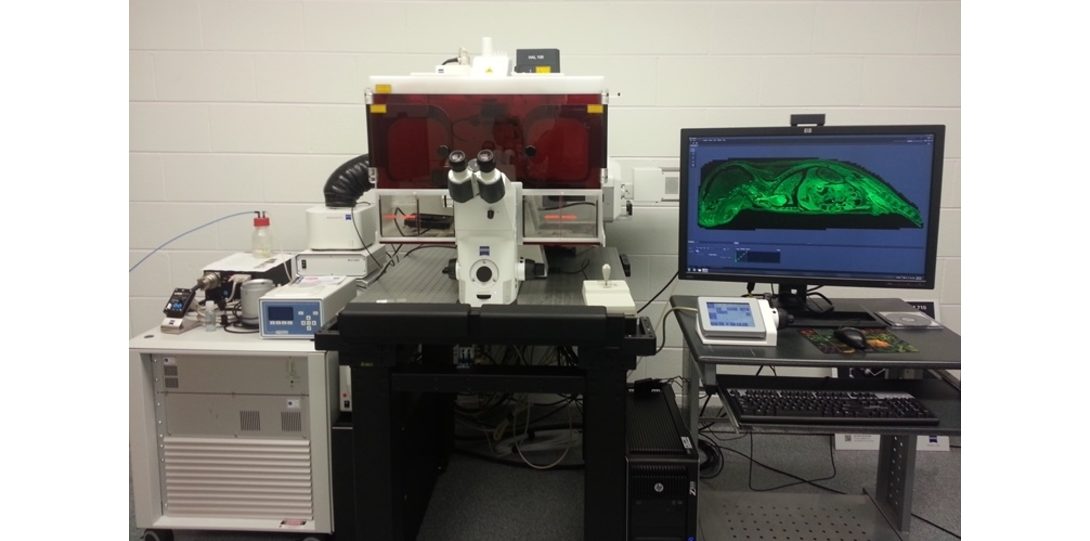
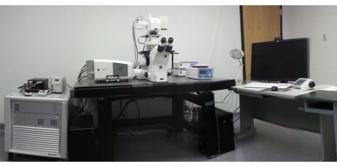
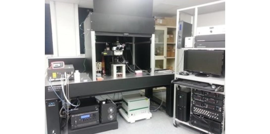
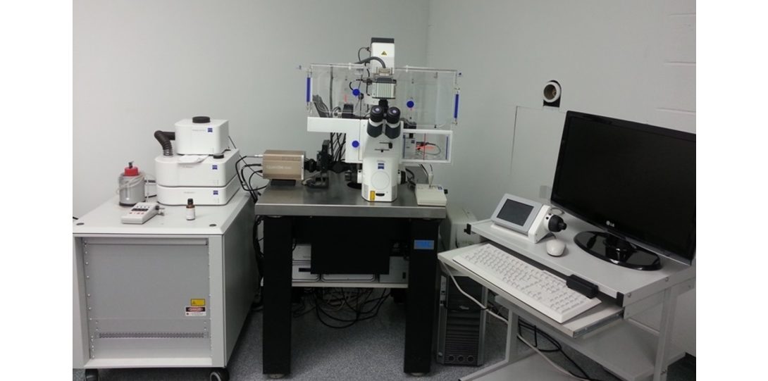
Image Processing Software at the FIC Heading link
Imaris Scientific 3D/4D Image Processing & Analysis Software
- One of the gold standards in Microscopy Image Analysis software packages
- Version 10.1 available on dedicated workstation (Puget Custom computer, 256GB RAM) now includes AI microscopy image analysis, stitching and deconvolution plugins.
MetaMorph Premier Image Analysis Software
- One of the most feature-rich analysis packages available for fluorescent microscopy
- Contains comprehensive tools for thresholding, filtering, and performing all manner of morphometry analyses
- On offline version of the software is available on a single PC workstation in the facility
- For a complete list of available modules, visit the MetaMorph Premier info page
Zeiss LSM Image Browser
- Free small standalone software for opening .czi, .lsm files
- Permits basic adjustments to prepare and export images for publication
- Available for Windows operating systems only
- A copy of the software can be found on the Zeiss product page.
Zeiss ZEN Blue and black off-line
- More extensive option for opening and altering Zeiss microscopy files
- Manage, measure, and export images with an interface matched to the ZEN software
- Available for Windows operating systems only
- Proceed to the software homepage for more information and a free download link of ZEN Lite (requires installation)
ImageJ and Fiji
- An open source, fully-customizable, NIH-developed image processing program
- Reads all major file formats (some may require additional plugins)
- Available for Windows, Mac OS, and Linux
- Visit the ImageJ and Fiji site for a full description, download link, and list of all plugins
Reservation Heading link
All of the instruments in active use within the FIC can be reserved via the online scheduler. Reservations are currently limited to 4 hours per day. (Special reservation guidelines apply to the Prairie In Vivo two-photon system, see instrumentation page) Users are not permitted to book an instrument for others if they will not be present during the allotted time. These guidelines are intended to avoid the monopolization of a single system by any individual laboratory.
Please use the FIC scheduler to reserve your microscope time.
To get information about payment sources, please click here.
Billing Heading link
Billing cycle is done on the monthly basis. On the 3rd of each month, the charges accumulated for the preceding month are sent to RRC finance for invoicing to the proper RRC account number.
Please note, Investigators from the Northwestern University and the University of Chicago are provided internal rates as all UIC users. All other users can access services but, at either external academic or external non-academic rate.
Rates and Instrument usage fees: Heading link
| By Price Groups | Rate |
|---|---|
| UIC (Internal) | 1x |
| Academic (Internal) CBC agreement with UC, Northwestern, Illinois Medical District | 1x |
| Other Academic | 1.599x |
| Other non-profit | 1.599x |
| Industrial | ~2x |
title Heading link
| Instrument | Rate |
|---|---|
| Confocals - 2 x 2 hours required | ~$380 |
| Motorized TIRF/Spinning Disk - 1 x 2 hours required | ~$190 total |
| Olympus VivaView Incubator Fluorescence Microscope | no training fee |
| Olympus BX51/IX70 Fluorescence Microscope | no training fee |
| Image analysis workstation | no setup fee |
| Bruker 2Photon microscope 3 x 2 hours required | ~$636 |
title Heading link
| Current Instrument | Rates (UIC internal rate, 1X) |
|---|---|
| Zeiss LSM 710 Confocal Microscope (BIG) | $45.00/hour |
| Zeiss LSM 710 Confocal Microscope (META) | $45.00/hour |
| Zeiss Laser TIRF Microscope | $45.00/hour |
| Zeiss Spinning Disk Confocal Microscope | $45.00/hour |
| Zeiss Widefield Microscope | $45.00/hour |
| Imaris Image Analysis Workstation | $20.00/hour |
| Olympus BX51/IX70 Fluorescence Microscope | $22.00/hour |
| Olympus VivaView Incubator Fluorescence Microscope (1-4 hours) | $22.00/hour |
| Bruker 2Photon Microscope (2P laser hours) | $56.00/hour |
| Assistance/Consultation | $50.00/hour |