Instruments
Instrumentation and services are available to all researchers inside and outside of the university.
Please check out the policies page (bottom) for information on charges.
Researchers from the Northwestern University, the University of Chicago and the Illinois Medical District are charged at internal rates as all UIC users. All other users can access services but at an external academic or external non-academic rate.
For regular users EMC will train you on the instruments you need for your research. Get in contact with the appropriate staff member to schedule training (Olivia Thomson - SEM (Hitachi S-3000N), Figen Seiler - Life Science TEM (JEOL JEM-1220) or Fengyuan Shi - Materials Science TEM (JEOL JEM-3010), Surface Science (XPS or Raman).
Users that need the high-resolution instruments (JEM-ARM200CF) must have sufficient previous microscopy experience on the JEOL JEM-3010 respectively before training on the high-resolution instruments. Once trained users can book instrument time using iLabs. For all other users, bookings should be made by e-mail or phone to the appropriate staff member (Figen Seiler for JEM-1220 TEM, Olivia Thomson for S-3000N VPSEM or JEOL IT500HR; Fengyuan Shi for all other instruments.)
EMC Staff have the final say as to whether a specimen can be looked at on an EMC instrument, and what instrument should be used for a specific specimen. It is our responsibility to maintain the instrument in a working state for all EMC users. If we believe a specimen to be potential damage to the vacuum system or optics we will refuse access or insist the specimen be looked at in a different instrument. Specimens that may be unsuitable include magnetic specimens, wet or water containing specimens and specimens that will evaporate in a vacuum. If you are unsure about the suitability of a specimen please ask!
Please check out our guide to the digital imaging ethics before publishing any work from EMC
All users are responsible for their own laboratory safety training to be current during each use of core facilities.
List of Instruments Heading link
| Instrument | Instrument Name | Location |
|---|---|---|
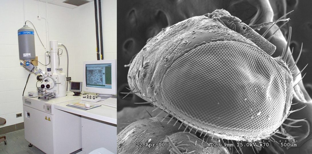
| Hitachi S-3000N | Variable Pressure Scanning Electron Microscope | Medical Sciences Building, E-5G |
| In-Situ Stages | Special Holders for Transmission Electron Microscopy | Science and Engineering South, 116A, 104B |
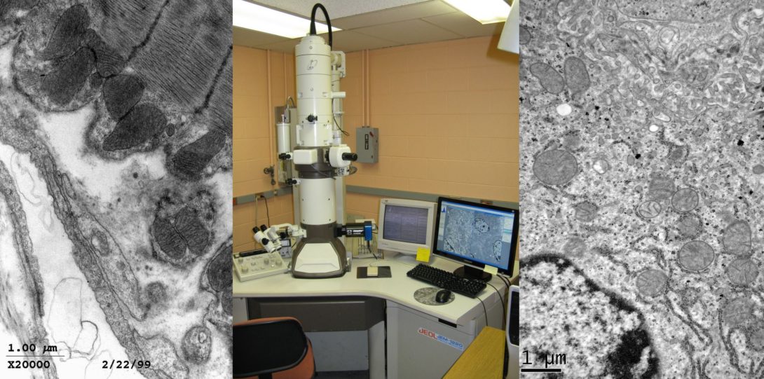
| JEOL JEM-1220 | Life Science Transmission Electron Microscope | Medical Sciences Building, E-5H |
| JEOL JEM-1400 Flash TEM | Life Science Transmission Electron Microscope | Medical Science Building, E-5 |
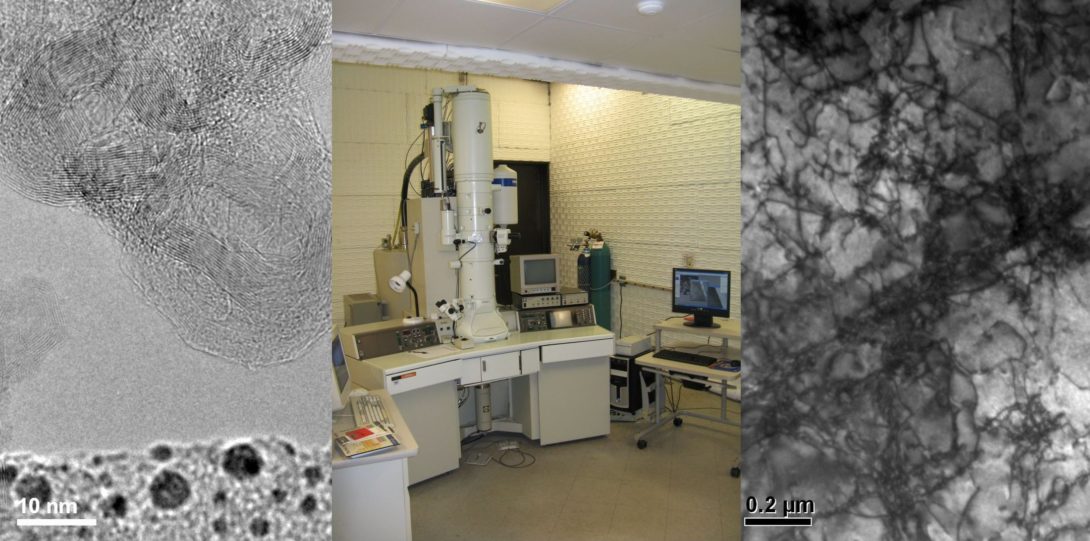
| JEOL JEM-3010 | Materials Science Transmission Electron Microscope | Science and Engineering South, 116C |
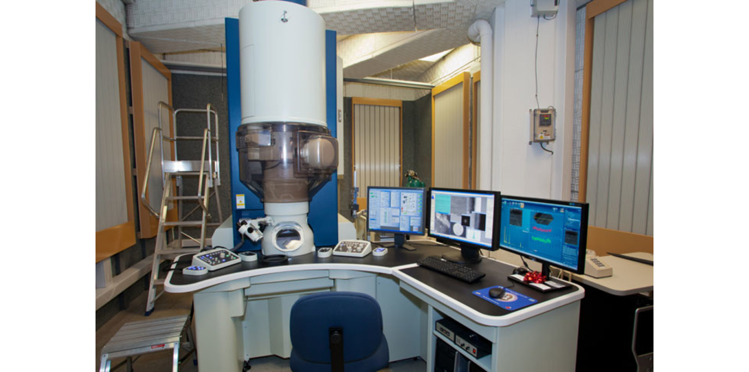
| JEOL JEM-ARM200CF | Aberration Corrected Cold Field Emission Scanning Transmission Electron Microscope | Science and Engineering South, 104A |
| JEOL JSM-IT500HR FESEM | Field Emission Scanning Electron Microscope | Science and Engineering South, 109C |
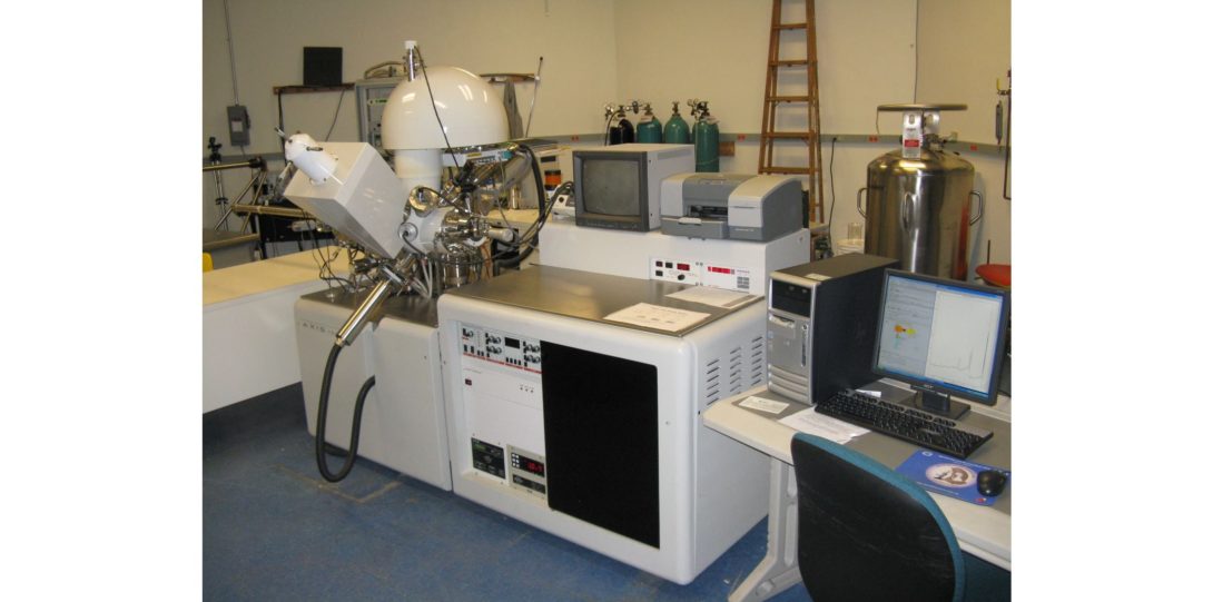
| Kratos AXIS-165 | X-Ray Photon Spectroscopy | Science and Engineering South, 109B |
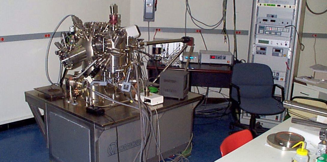
| Omicron VT-SPM | Ultra-High Vacuum Scanning Probe Microscope | Science and Engineering South, 109B |
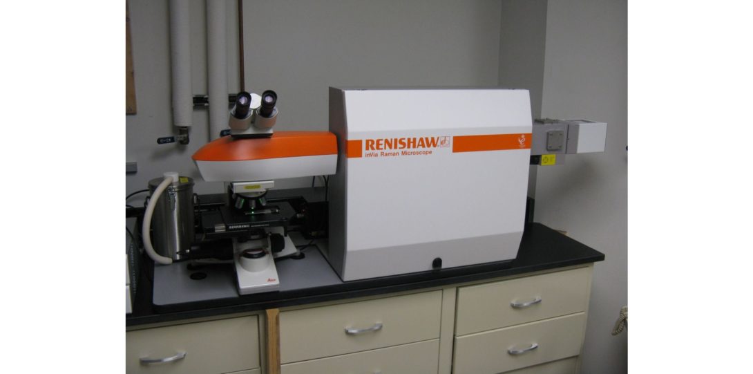
| Renishaw inVia Reflex | Micro-Raman Spectrometer | Science and Engineering South, 116B |
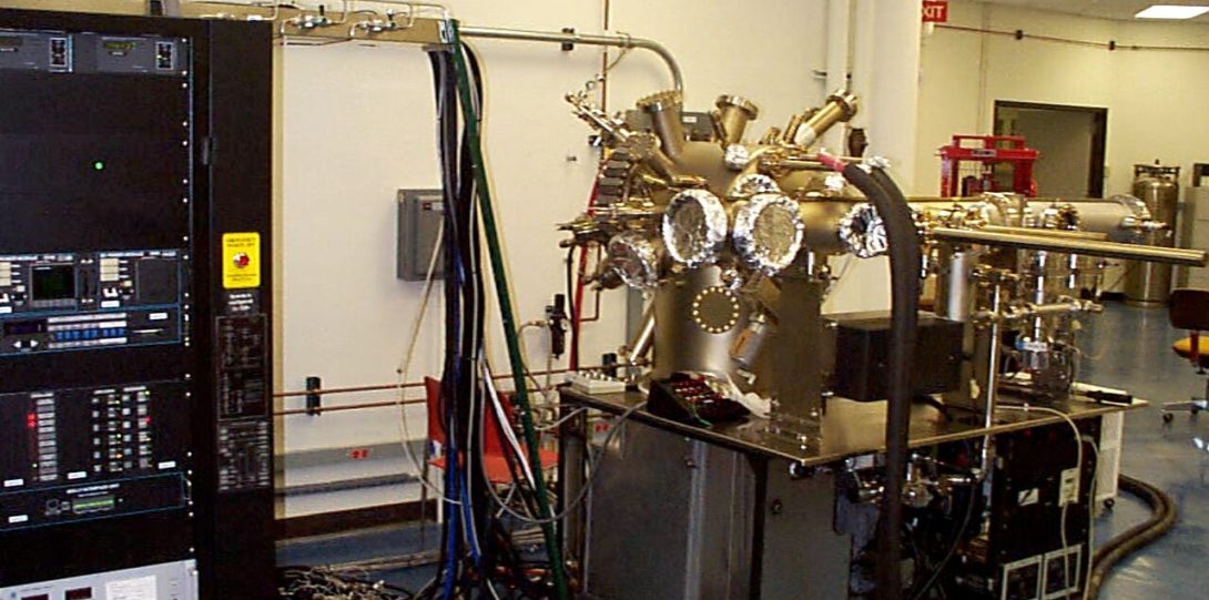
| Veeco GEN II | Oxide Molecular Beam Epitaxy | Science and Engineering South, 109B |
Preparation Laboratory EMS-E Heading link
Location: Science and Engineering South, 116A
Description
There are many ways of preparing specimens for electron microscopy. The EMS staff are available to help investigators determine the best way of getting the information they need from their specimens. The following is intended only as a general guideline.
All EMS TEMs take standard size specimens (3mm in diameter). The maximum thickness of the specimen depends on the density of the material, the accelerating voltage of the microscope and the resolution of the analysis to be carried out. The EMS has extensive specimen preparation facilities to convert bulk material into transmission thin samples of the correct diameter. Specimens can be either self-supporting (i.e. the whole specimen consists of one material) or supported on a grid or a slotted 3mm washer.
Semiconductor, Superconductor and Metal specimens typically are polished down to 100-200 micrometer, a 3mm disc is cut from the material, then the central region is pre-thinned to a few micrometers before final thinning, to perforation, is carried out by Ion Beam Thinning, Electropolishing or Chemical Thinning. Another approach is to slice and mount on a grid before polishing down and then final thinning to perforation.
Catalyst or Ceramic specimens can often be crushed, dispersed in alcohol and a drop of liquid placed onto a grid supporting an amorphous carbon film.
Polymer specimens are typically prepared using an ultra-microtome to cut slices less than 100nm thick which can then be supported on a grid.
Equipment available in the EMS-E prep lab includes:-
- Diamond saw for slicing (South Bay 650)
- Ultrasonic Cutter, Disc Cutter and Rotary Slurry Cutter for preparing 3mm discs (South Bay 380, 310 and 360)
- Polishing System (Allied High Tech Multiprep)
- Polishing Wheels (South Bay 900 and 910T)
- Tripod Polishers (South Bay 590W) and Disc grinders (Gatan 623)
- Dimpler/ Polisher (South Bay 515)
- Plasma Cleaner/ Etcher (South Bay PC150)
- Twin Jet Electropolisher (Fischione 110)
- Ion Mills (Fischione 1010 and M1050)
- Ultramicrotome (Leica UCT)
- Chemical Thinning Apparatus
- Ultrasonic Cleaner (Branson 1510)
- Reflection Stereomicroscope (Fisher Scientific Stereomaster)
- Transmission Polarizing Microscope (Nikon Labophot-Pol)
- Transmission Light Microscopes (Leica ATC2000, AO Scientific Instruments One-hundred Microstar, Bausch & Lomb)
Preparation Laboratory EMS-W Heading link
Location: Medical Sciences Building, E-5B
Description
There are many ways of preparing specimens for electron microscopy. The EMS staff are available to help investigators determine the best way of getting the information they need from their specimens. The following is intended only as a general guideline.
The size of the SEM specimen depends on which instrument you need for your analysis. The Hitachi S-3000N is able to accept specimens up to 150mm in diameter (although parts of the specimen cannot be accessed). The JEOL JSM-6320F can accept specimens up to 30mm in diameter and 10mm high. In general, however, the amount of material should be kept as small as possible, especially if it is non-conducting. Conducting specimens can be imaged in both SEMs without any further specimen preparation. Non-conducting specimens may need coating with a conducting film in order to reduce charging at high vacuum in all microscopes. The EMS is able to coat with either carbon, platinum/gold, platinum/palladium or, for high resolution imaging, chromium. In low vacuum mode (S-3000N only) it is not necessary to coat any specimen and some non-conducting specimens can be imaged without charging at low voltage (S-3000N, JSM-6320F).
All EMS TEMs take standard size specimens (3mm in diameter). The maximum thickness of the specimen depends on the density of the specimen. The EMS has extensive specimen preparation facilities to convert bulk material into transmission thin samples of the correct diameter. Life Science Specimens are supported on a grid or a slotted 3mm washer. Life Science specimens are typically prepared using an ultra-microtome to cut slices less than 100nm thick which can then be supported on a grid.
All life science specimens (for SEM or TEM) need to be processed to remove the water from the specimen and replace it with a plastic resin without introducing artifacts. Typically, for TEM, this is a multistage process involving
- Fixation – to halt degradation of tissue
- Dehydration – to replace water with ethanol and then a transitional solvent
- Infiltration – to impregnate the specimen with resin
- Embedding – to enclose the resin impregnated specimen in more resin
- Ultramicrotomy – to cut transmission thin slices from the specimen
- Staining – to increase the contrast of the images obtained by staining parts of the structure with a heavy metal stain (eg uranyl acetate)
Equipment available in the EMS-W prep lab includes:-
- Sputter coaters for depositing metal films (Cressington 208HR (Cr) and Polaron E5100 (Pt/Pd & Au/Pt)).
- Tissue Rotator (EMS), Centrifuge (Spectrafuge 6C)
- various Ovens
- Glass Knife Maker (LKB 7801B)
- Ultramicrotomes (Leica Ultracut UCT and Reichart Ultracut E)
Ultrafast Electron Microscope Heading link
Location: Science and Engineering South, 119
Description
The Ultrafast Physics Group in conjunction with RRC-E are developing an Ultrafast Electron Microscope. This instrument aims to combine the high spatial resolution of electron microscopy with the ultrafast (sub-picosecond) time resolution possible using ultrashort pulse laser systems.
The aim of this project is to develop a functional prototype UEM with a space time resolution of less than 1nm-ps.
At present most work has centered on the development of the ultrafast electron source, eventually this will be mated to the bottom half of an existing TEM and made available to researchers at UIC.